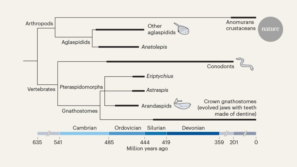
Synchrotron Tomography of Late-Cambrian Fossil Fragments. IV. Anatolepis and its Vertebrate Affine
To test the vertebrate affinity of Anatolepis, we first deployed high-resolution synchrotron tomography on late-Cambrian fossil fragments. The canals are connected to the cavities by a thin layer of elysian fibroblasts. The tubercles connect to a central cavity that extends to the ventral surface and dorsally attenuates into multiple large-calibre tubules (Fig. 1h). These features were previously interpreted as a pulp cavity and dentine tubules, respectively, in scanning electron micrographs3. Although Anatolepis’ tubules were difficult to segment because of partial collapse due to acid preparation, tubules can be seen to have a distinct flared arrow-shaped end in the virtual transverse section (Fig. 1b). The center of thetubercle contains a few peripheral tubules that surround the edges. Notably, the peripheral tubules had been observed previously in scanning electron micrographs3 but interpreted as the natural odontode edge. Externally, Anatolepis fragments are marked by rounded tubercles that are interspersed and non-overlapping (Fig. 1a and Extended Data Figs. 1 and 2). Anatolepis’ tubercles vary subtly in morphology and are situated within the thin basal tissue; the tubercles also extend above the basal tissue that is marked externally by several pore openings (Fig. 1 and Extended Data Figs. 1 and 2). The tubercles on the opposing side are marked by a series of grooves that match the direction in which they are located on the ventral side. We used this distinct internal and external morphology to match our samples with previously described specimens of Anatolepis3,5.
In comparison to Anatolepis, high-resolution phase-contrast scans of coeval aglaspidid arthropods were obtained. There is also phosphatic 41,42, 43 in the exoskeleton of aglaspidids. Aglaspidid cuticles have a homogeneous lamellar structure that is interrupted by vertical pore canals like those described in Anatolepis3 (Fig. 1 and Extended Data Figs. 1–3). Some of these pore canals flare internally into teardrop-shaped cuticular organs, which vary in size through the specimen and are similar to the Anatolepis specimens (Fig. 1c,d and Extended Data Figs. 1e,f and 2h). In Aglaspis? We resolve horizontal canals, as well as the vertical canals and cuticular organs. However, this feature was not seen in all scanned aglaspidid cuticle samples and is notably absent from thicker cuticular regions such as portions of the tail spine (Extended Data Fig. 3a,d The virtual sections show partially in filled central cavities like the ones in Anatolepis. The central tubules have a distinct arrow-shaped cavity and are capped by a cone. Each central tubule is surrounded by a hypermineralized layer that is then surrounded by a hollow cavity, in a tube-in-tube formation that is not seen in vertebrate dentine (Fig. 2e,f). The tubules circumferentially diffuse from the central cavity, and the hollow cavities merge and form a honeycomb structure that is hollow (Fig. 2c,e). Additionally, there is a set of tubules that are peripheral to the central tubules; these are simpler in morphology, lacking both the flared end and mineralized coating (Figs. 1h and 2e). Externally, aglaspidid cuticles have diverse tubercles that vary in size and morphology but are generally rounded, with a corresponding dimple on the underside, the same characters that define Anatolepis (Fig. 1a,e). The external morphology, scans and subsequent segmentation of several aglaspidid cuticle fragments revealed microanatomy with all of the hallmarks of Anatolepis, in having a lamellar basal tissue perforated by vertical and horizontal canals, a central cavity, teardrop-shaped cuticular organs and characteristic arrow-shaped tubules. We concluded that Anatolepis is not a species of mammal, but rather a species of arthropod. As this taxon was the only putative stem-gnathostome with dentine from the Cambrian, this identification pushes the earliest definitive occurrence of the clade 40 million years into the Middle Ordovician6.
