Fold Changes in Level 4 Neuron Subtypes after SCI and their Correlation to Post-Injury Recovery for Vsx2ON Neurons
a, Heatmap showing fold changes for all genes differentially expressed after SCI in at least one level 4 neuron subtype at 1 day, top, and 1 month, bottom, after injury. Patterns of differential expression are broadly conserved at 1 day, but more subtype-specific at 1 month. A heat map shows the fold changes for genes with broadly conserved patterns of differential expression right after an injury. c, Heatmap showing coefficients estimated by linear mixed models within each neuron subtype for up- or downregulation of the early-conserved neuronal module over the injury timecourse. d, Heatmap showing coefficients estimated by linear mixed models within each neuron subtype for selected GO term modules with broadly conserved patterns of up- or downregulation across level 4 neuron subtypes at 1 day post-injury. e, Dot plot showing median expression of the circuit reorganization module in each level 4 neuron subtype across timepoints. The size and point color of the circuit reorganization module show it’s expression. The boxes show the expression of the axon development, left, and dendrite development. There is a circuit reorganization module that is within level 4 of the uninjured spine. Vsx2-expressing neurons display the highest expression of the circuit reorganization module in the uninjured spinal cord. The growth factor Gdnf is shown to be expression across levels 1 to 4 of the neuron taxonomy. The mean expression of each neuron subtypes is displayed by the point color and the point size. i, Scatterplots comparing basal expression of the circuit reorganization module in the uninjured spinal cord, x-axis, with the SCI-induced upregulation of this module at each timepoint after injury, y-axis, for each level 4 neuron subtype. The text shows the correlation. Basal and induced expression of the circuit reorganization module is maximally correlated at 1 month post-injury, coinciding with the temporal window of opportunity for natural recovery after SCI. j, Timeline of Vsx2ON neuron diphtheria toxin ablation experiments. Animals received injection of AAVs expressing DTR two weeks before crush. Animals were given daily injections for 7 days at eight weeks. Tissue was collected to evaluate the Kinematics. Histology verification of Vsx2ON neuron surgery in the lower thoracic region. Above and below the level of the crush SCI, there is loss of Vsx2ON neurons. The number of Vsx2ON neuron found in each animal is shown in a bar graph. In the Vsx2ON experiment, locomotor performance can be quantified by the number of mice per group.
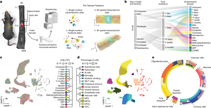
The spinal cord, like the brain, is made of delicate tissue that is isolated from the body’s immune system by physical barriers that restrict the entry of immune cells. When the brain is damaged, the body’s immune system responds by attacking the injury site. It can make injury worse by making it infections and impair healing. The researchers showed that the influx peaks between 7 and 14 days after injury.
Atlas of Genes to Support Old Mice Against SCI: A Study Based on a Cell-Biology and Machine-Induced Atlas
Anderson and his colleagues used machine-learning algorithms to build the atlas by mapping data from RNA sequencing and other cell-biology techniques. They described the work in a Nature paper published today1 and have made the entire atlas available through an online platform.
When injected into old mice before they had SCI, the treatment increased the number of border- forming astrocytes, reduced the number of harmful immune cells and restored the integrity of the blood–spinal cord barrier. As a result, treated mice had smaller and more contained spinal-cord lesions and recovered their ability to walk just as well as young mice that experienced similar injuries.
It is possible to see that the barriers form very robustly in young animals but are not functional in old mice, according to Anderson.
Older mice had larger tumors with more extensive loss of nerve cells and increased invasion of immune cells. Their ability to recover from SCI was also reduced, leading to functional impairments and paralysis.
The scientists designed a therapy to promote wound repair in older mice using insights from the atlas. They used a virus to deliver genes programmed to express three growth factors. These proteins can boost the growth of astrocytes and cells that form blood–spinal cord barrier.
The researchers say the gene-therapy component of their study provides proof of principle, but caution that more work is needed before this approach could potentially benefit people with similar injuries.
Gene therapy effects can last for years, but one challenge will be controlling the duration. Timothy O’Shea is a medical engineer at Boston University in Massachusetts.
To administer such treatments, it’s important to determine the best time to do so. “Those things are still the kind of caveats that I think would need to be worked out from a therapeutic standpoint,” says O’Shea.